VinBrain Case Study: AI Assists in Follow-up of Pneumothorax Patients


Ferrum Health partners with best-in-class AI providers offering solutions designed to reduce radiology workload and improve patient care.
VinBrain Case Study: AI Assists in Follow-up of Pneumothroax Patients
Introduction
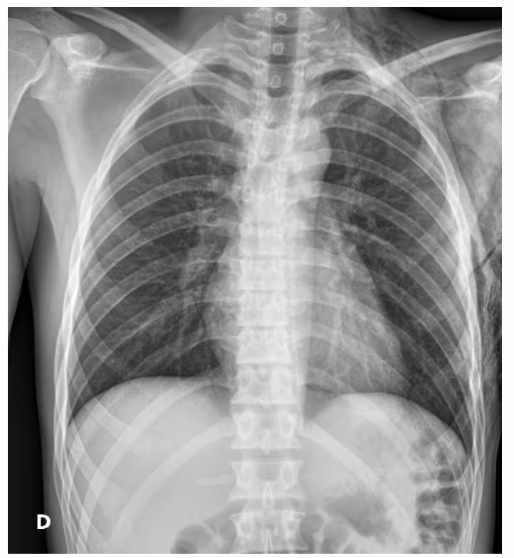
To prevent quick progression of small pneumothorax to a large-size pneumothorax, chest x-rays must be repeated within 3-6 hours after admission for patients on observation. Spontaneous hemopneumothorax must be considered during this period. Imaging evaluation should be done within 12-48 hours after discharge. Admission observation is recommended for patients without reliable follow-ups to prevent the condition from worsening.
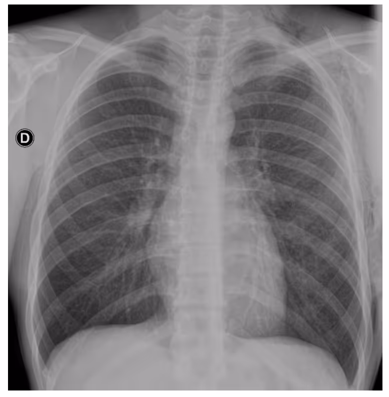
Clinical Case
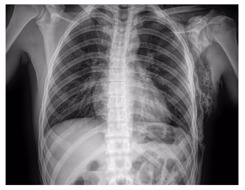
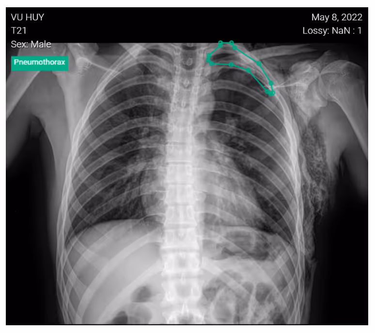
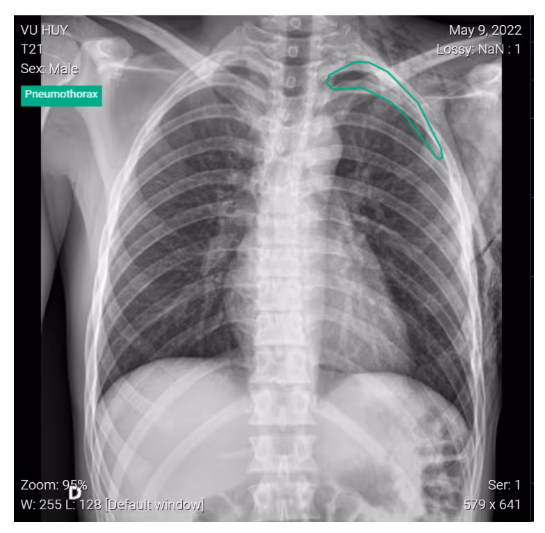
- A 20-year-old male presented with a stab wound to the left hemithorax, and crepitus of the chest was detected upon physical examination.
- Image findings:
- Chest radiograph revealed a left pneumothorax, marked by the visible left pleural edge
- No shift of the mediastinum
- Extensive subcutaneous emphysema over the left chest wall
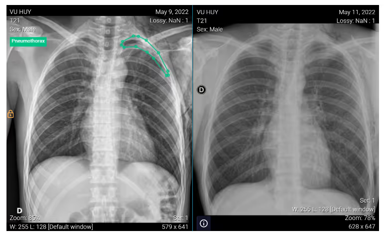
Conclusion
DrAid for Pneumothorax helps to compare side-by-side images for follow-up in pneumothorax patients. This aided reading allows doctors to create a suitable treatment plan for the patient.Interested in deploying DrAid at your health facility?