Customer stories
VinBrain Case Study: Quick Detection of Acute Cases - Pneumothorax
June 30, 2022


Ferrum Health partners with best-in-class AI providers offering solutions designed to reduce radiology workload and improve patient care.VinBrain Case Study: Quick Detection of Acute Cases - PneumothoraxIntroductionA patient presented to the emergency department with the acute onset of shortness of breath and chest discomfort. It is difficult for physicians to diagnose with no significant medical history.Clinical Case
- A 20-year-old male came to the emergency room complaining of shortness of breath and chest pain.
- The patient was healthy with no significant medical history
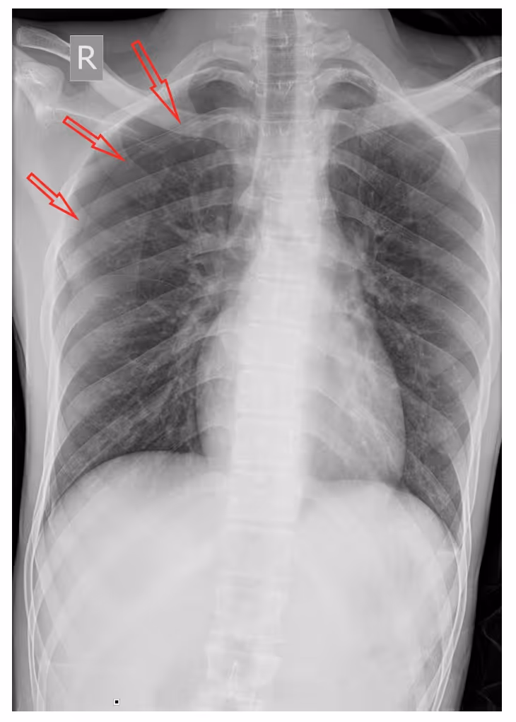
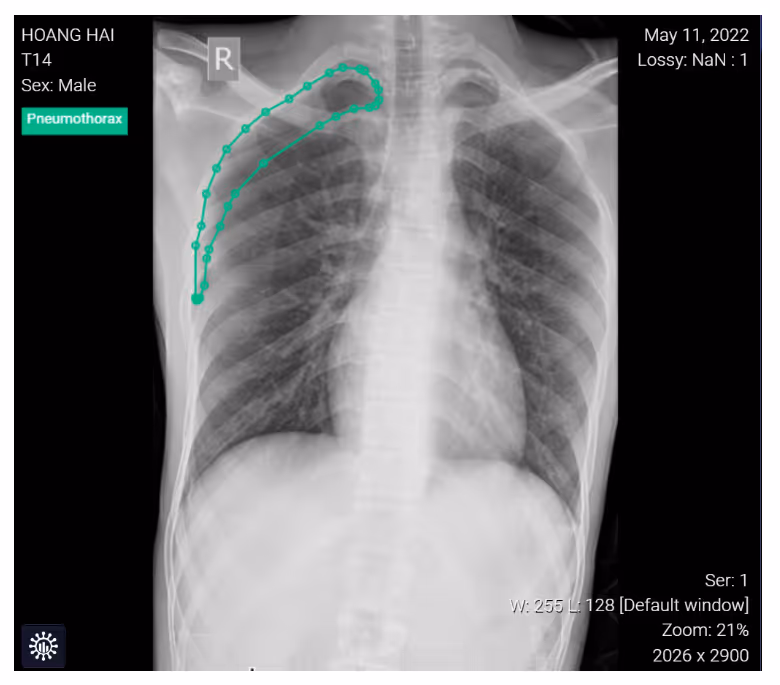
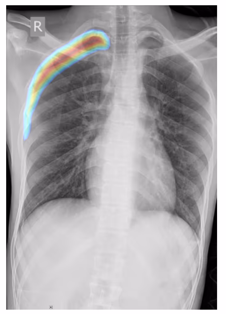
- A frontal chest radiographic image highlights the classic appearance of right-sided pneumothorax with a readily apparent visceral pleural line as seen without distal lung markings

ConclusionPhysicians can use DrAid for re-checking diagnostic results, thereby supporting the quality and efficiency of doctor performance.Interested in deploying DrAid at your health facility?Contact the Ferrum Health team to learn more.