Customer stories
VinBrain Case Study: Accurate Analysis for ICU Patients - Pneumothorax
July 14, 2022


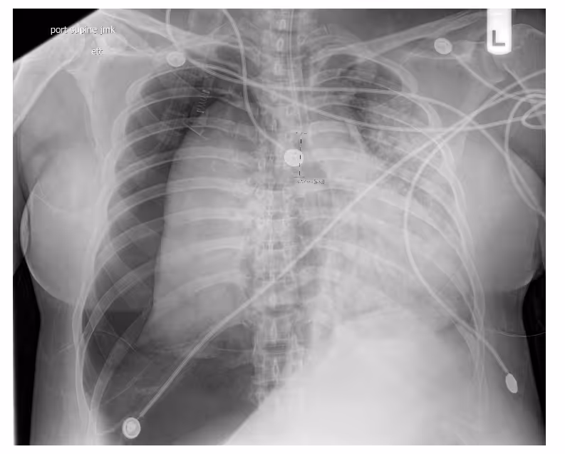
Ferrum Health partners with best-in-class AI providers offering solutions designed to reduce radiology workload and improve patient care.VinBrain Case Study: Accurate Analysis for ICU Patients - PneumothoraxIntroductionICU patients often have chest x-ray images taken at the bedside with a portable x-ray machine while many medical devices are placed on their bodies. This makes diagnosis more complex than a common patient.Clinical Case
- A 40-year-old female patient with post endotracheal tube placement with known COViD pneumonia
- Image findings:
- Interval placement of endotracheal tube terminating 3 cm from the carina.
- Large right tension pneumothorax and scattered lucencies are seen.
- The cardiomediastial silhouette is shifted into the left hemithorax, and the vasculature is obscured.
- Complete collapse of the right lung.
- Diffuse patchy airspace opacities in the left hemithorax
- No pleural, osseous, or soft tissue abnormalities
- The patient has many medical devices placed on their body, and more than one abnormal finding appears on the lungs.
- The patient was diagnosed with sizeable right tension pneumothorax, with a complete right lung collapse and a leftward shift of the cardiac mediastinal silhouette.

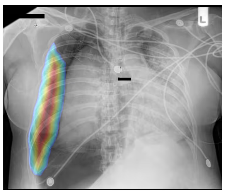
ConclusionDrAid for Pneumothorax was able to accurately analyze the suspected pneumothorax on the x-ray images of the ICU patient with many overlapping findings.Interested in deploying DrAid at your health facility?Contact the Ferrum Health team to learn more.